Blogger Widgets

...

TRANSLATE AS YOU LIKE

April 21, 2012

Post-Marketing Pharmacovigilance for a New Drug

Pharmacovigilance is the scientific study of medicinal products intended to treat a particular disease or indication for humans. It aims to establish drug safety profiles; knowledge about efficacy; metabolism and excretion; and other important clinical information. Within the EU and elsewhere, the regulatory authorities require all new medicinal products to have successfully completed a series of clinical trails before being granted a Marketing Authorisation which allows them to go on sale. This article explains some key points about the need for clinical trials and studies after the drug reached the market.
Pre-Marketing Clinical Studies Have Their Limitations
During the phase before a Marketing Authorisation is granted, clinical studies could have involved up to 10,000 patients. However, this is not always the case: it is also possible for the study size to have been much smaller. For example, ‘orphan drugs’ are those intended to treat relatively rare diseases. There may be cases where these drugs have been studied using smaller trials, with perhaps only dozens to hundreds of patients taking part.
Although pre-marketing trials must have been conducted in an extremely rigorous manner in order to meet EU drug safety regulations, they nonetheless have their limitations. The number of patients involved in even the largest trial means that uncommon and rare adverse reactions are statistically unlikely to have been detected with any certainty. The trials are able to produce important data but for example, at 5% probability level the absence of an adverse reaction within a study of 10,000 patients produces a true rate of occurrence of less than 1 in 3,333.
Pre-Marketing Trials May Not Include All Patient Groups
It could be that clinical trials did not include all the patient groups for whom the drug would be prescribed to. An example here could be studies involving patients with a maximum age of 60 years participating in clinical trials before the drug goes on sale, and subsequent prescribing to older patients once it reaches the market. There are a number of issues which could be relevant within this example. This patient group could be taking numerous other prescribed medications; they could present variations of metabolism and body mass index in comparison to younger patients; and they could present with a heightened sensitivity to certain types of medication. There could be issues around food intake, for example, if these patients are unable to follow a regularly timed meal schedule for any reason. Although there may have been some investigation into issues of polypharmacy, metabolism, the effects of food intake and so on during the pre-marketing studies, it could be that this particular patient group were not included in those studies.
Once the drug has been placed onto the market, the first 500,000 to 1,000,000 patients who take the new drug represent the first large scale safety test. Nurses, doctors and pharmacists around the world report suspected adverse reactions as spontaneous reports. They may do so via the regulatory agencies; national pharmacovigilance centres; or directly to the pharmaceutical companies. If a pharmaceutical company receives one of these reports, it must report it to the regulatory authorities. This will be generally within a 15 day timescale, subject to national and international laws.
Please note it is impossible to provide an exhaustive description of this topic within this short article; readers are therefore advised that this information cannot be considered as any type of professional advice.
by
Akshaya Srikanth
Pharm.D Resident 
India

April 19, 2012

Lymphatic Filariasis

Enlarged, red and painful legs, thick and creamy urine, monstrously large limbs or scrotums, fits of asthma... lymphatic filariasis manifests itself in many different forms depending on the individual. These symptoms are due to the obstruction of lower lymphatic vessels by worms of the genus filaria, transmitted to man by the stings of diverse species of mosquitoes.
At the beginning of the 20the Century, the essential elements of the fundamental data concerning the disease are known. The Colonial Health Service determines the geographical distribution of this endemic which, in the zone under French influence, affects dozens of millions of individuals. These physicians participate in the study of the acute manifestations of the disease. The considerable longevity of the worms (more than 15 years) explains the chronic evolution of the ailment with various manifestations, of which the most spectacular is Elephantiasis.
Up to 1947, no medical treatment is efficacious. Only surgery can relieve those who suffer from Elephantiasis. Techniques, which vary in the course of the century, are proposed one after the other and put into practice. In spite of the precarious conditions, success is often remarkable.
The advent of anti-filaria remedies permits the undertaking, notably in Polynesia, of the first chemoprophylaxis mass campaigns.
In 1862, in Paris, Demarquay discovers embryos of filaria in the blood of a Cuban. The same phenomenon is observed by Wücherer in Brazil in 1866, then in India and Guadeloupe... but it is Bancroft who finds the adult filaria in Australia in 1876. It is named bancroftian filariasis and, later, Wuchereria bancrofti. At the same time, P. Manson designates the mosquito as the transmitting agent of the parasite and Low, in 1900, demonstrates that this transmission is due to the stinging of the insect.
Later, three lymphatic filaria are identified but only Wuchereria bancrofti, a cosmopolitan parasite which only contaminates humans, is to be found in the French colonial domain.
Filaria are thin round worms a few centimetres long. Doted with remarkable longevity, the female, in the course of 15 years, emits millions of larvae (microfilaria). These manifest relatively little aggressiveness and are incapable of evolving into the adult stage in their environment, as passage through mosquitoes is essential for that. At night, they leave the lymphatic vessels to enter the blood stream. At sunrise, as Manson showed in 1877, they are no longer there.
When absorbing their repast, nocturnal mosquitoes imbibe microfilaria together with the blood of a patient.
Adult filaria abide exclusively in the lower lymphatic vessels of the abdomen.
Microfilaria of Bancroft seen through a microscope
In Indochina as in the Indian Trading Posts, the physicians of the Colonial Health Service have reported the disease since 1908 in Cochin China (Noc*), in Tonkin (Mathis* and Léger*). The rate of infection is estimated as 15 % in the population of Saigon.
The Pacific Islands are greatly affected (Brochard*). In 1940, 60 % of the population is smitten.
In the West Indies, lymphatic filariasis has been identified since 1907 (Dufougéré*). In 1914, Léger* and Gallen* report the presence of the parasite in 15 % of the population. The situation is the same in Guyana.
Geographical distribution of lymphatic filariasis
In sub-Saharan Africa, Thiroux* in 1912 and Léger in 1913 show the widespread nature of the endemic in the countries of French West Africa (AOF) and French Equatorial Africa (AEF).
In the Indian Ocean, it is the same as in the other territories, notably in Reunion Island (Thiroux*) and the Comoro Islands (Rouffiandis* 1910). There more than 80 % of the population is infected.
The rates of infection are very much superior to the number of patients who show symptoms of filariasis. "Healthy carriers" are numerous and contribute towards the perpetuation of the endemic.
ACUTE MANIFESTATIONS
A few months after contamination, there follow the first acute manifestations. Their repetitive character attracts attention. It is a question of very high fever accompanied by pains in the scrotum and the inguinal region or acute repetitive lymphangitis of the limbs. The leg is swollen and painful, the skin red and glistening. In the groin, the lymph glands are sensitive.
Acute lymphangitis of the leg
The explanation of these troubles opposes, for a long time, those who favour a microbic origin and those who advocate a filarial origin. The extremely inflammatory aspect leads to the search for a microbic component : besides the streptococcus, some think there is the intervention of a particular germ called the lymphococcus by Dufougéré* and the dermococcus by Le Dantec*. The filaria theory, upheld by Manson, is confirmed by the experience of American military physicians in the Pacific, between 1947 and 1950 : the use of recently-discovered anti-filaria remedies cures their patients afflicted with lymphangitis.
The explanation of the symptoms of filariasis is arrived at thanks to lymphography, x-rays of the opacification of the lymphatic vessels. The obstruction of the vessels by clusters of adult worms, often dead, is responsible for most of the acute manifestations, in particular adenitis and oedemas, which can become more complicated in the case of a microbic superinfection.
The colonial physicians make a great contributions towards the recognition and study of this vascular obstruction, notably in Dakar, Dejou* in 1952, Carayon* in 1962 and Datchary* in 1963. Nowadays, besides the progress in the immunological diagnosis of the disease, non-interventional methods (lymphoscintigraphy and ultrasonography) permit the early detection of lower lymphatic obstructions. When precociously administered, the medical treatment proves to be efficacious.
CHRONIC EVOLUTION - ELEPHANTIASIS
Elephantiasis of the leg 
The repeated pressure of acute lymphangitis brings about the fibrous thickening of the skin and the subcutaneous tissue which, at length, can result in monstrous deformations called "Elephantiasis".

Elephantiasis of the foot
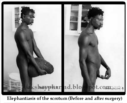
The parts of the body in which they usually occur are the legs, the scrotum and sometimes the penis, the vulva, the breasts... The lower limbs can look like the paws of a pachyderm. As for scrotums, becoming huge, they could weigh 30 to 40 kg, so that walking becomes impossible and the use of a wheelbarrow is obligatory for getting about. The penis disappears, embedded in the mass of flesh, but it remains unharmed as are the testicles too. Elephantiasis is here the consequence of a definitive obstruction of the lymphatic vessels, as lymphography confirms.
Lymphatic vessel walls are narrow and fragile and their distension in case of obliteration could end up in their rupture and fistulation. Chyle, abdominal lymph made creamy by fat globules from the digestive tract, then flows in the peritoneum (chyloperitoneum), in the intestine (chylous diarrhoea) or in the urinary tracts (chyluria).
THE TREATMENT OF LYMPHATIC FILARIASIS
Before the Second World War, no medical treatment yields satisfactory results : iodides, mercurial and arsenical derivatives, anti-streptococcal serum, electro and radiotherapies. Only surgery reduces distressing disabilities of the limbs and the genital organs by ablation of the fibrous mass in order to make the patient once again agile and virile.
Lemoine* is the first to practise "certain palliative interventions" in Tahiti in 1910. He sets about performing vast cutaneous resections called "melon slicing" ("en tranches de melon"). Other techniques are attempted but soon abandoned, such as external draining of the lymph or lympho-venous anastomosis...
In what concerns the scrotum, it is a matter of excising the skin after having extricated the testicles and the penis and then detaching the fibrous mass in one block. A primary obstacle is the volume of these scrotal masses, sometimes huge. The difficulty stimulates the imagination of the surgeons. Some of them suspend this mass from a pulley attached to the ceiling. Guyomarc'h*, about 1910, in French Equatorial Africa (AEF), begins the operation with a large incision in the middle which separates the mass into two symmetric parts. Then he extracts the testicles, the penis... It is the epoch of heroism : the patient is on a stretcher, the operation lasts about thirty minutes. The patient is glad to see his penis once again and to be able to resume sexual activity. Continued by Bernard* in 1911 and by Ouzilleau*, this procedure is used for a long time. The latter writes in 1913: "We have had only four failures in 182 operations". Unfortunately, the recurrence of the ailment is the rule, sooner or later.
After the Second World War, agreement is reached on well-codified techniques to be used in treating elephantiasis of the limbs. Servelle's techniques or those of Gibson and Touch are used by the surgeons of the Colonial Health Service.
To remedy filarial chyluria, the resection of lymph vessels of the affected kidney, proposed in 1964 by L. Léger, is used with success, particularly in Tahiti, by Fouques*, Huet*, Montangerand* and Roch* in 1967.
MASS ANTI-FILARIA CAMPAIGNS
They only become possible after the discovery of Notezine and they have two aspects :
Several varieties of Elephantiasis
- The fight against mosquito vectors and their breeding grounds, already undertaken in the combat against Malaria, may explain why today the filarial endemic with its spectacular examples of Elephantiasis is on the decline.

- Chemoprophylaxis, reserved for regions where filarial endemics rage. The whole population may receive Notezine (sometimes incorporated into kitchen salt) during 12 days every three months. Another procedure consists of taking the remedy every two weeks. In 1949, Laigret* begins a campaign in Polynesia and notes, in the population, a great reduction in the density of microfilaria in the blood.
These mass campaigns remain rare and are limited, for, in general, filariasis is not considered a priority. In 1998, the WHO, in a resolution of the General Assembly, demands member states to classify it as a public health problem.
Source: ASNOW
by
Akshaya Srikanth
Pharm.D Intern
Hyderabad, India

April 18, 2012

How Serotonin Reuptake Inhibitors Work: Understanding More Fundamental Mechanisms of Action

By stimulating microRNA miR-16 in the midline serotonergic raphe, fluoxetine initiates signaling cascades that lead to hippocampal neurogenesis.
Several lines of evidence suggest that in adults, antidepressant therapies enhance neurogenesis in the hippocampus, but how this process occurs has been unclear. These researchers studied the effects of fluoxetine in mice and in humans. They worked out several pathways that begin with the stimulation by fluoxetine of the microRNA miR-16 in serotonergic neurons in raphe and ultimately result in hippocampal neurogenesis.
In a series of experiments in mice, fluoxetine activated raphe miR-16, which decreased raphe levels of the serotonin reuptake transporter (SERT). In turn, these events directly caused brain-derived neurotropic factor (BDNF) and two other signaling molecules to act on the hippocampus. Indirectly, the same events resulted in release of another protein from the raphe nuclei, S100β, which in turn stimulated the locus coeruleus to induce SERT and secrete serotonin. Both the direct and indirect pathways caused decreases in hippocampal miR-16, which sequentially led to increases in both hippocampal SERT and the bcl-2 protein (which promotes neurotrophic function), which in turn stimulated neurogenesis. In nine patients with major depression, 12-week fluoxetine treatment increased levels of the three signaling molecules in cerebrospinal fluid. The interventions were accompanied by improvements in several mouse models of depression, as well as in the patients.
My Comment: These findings draw together several seemingly unconnected lines of research. The authors identify miR-16 as a "missing link" between serotonin reuptake inhibitor treatment and hippocampal neurogenesis and as a "micromanager" of the intervening changes in the raphe nucleus, locus coeruleus, serotonin receptor transporter, serotonin secretion, and hippocampal neurogenesis. The processes appear to work through the cooperative and integrated activities of several signaling molecules. Further clarification of these pathways may help refine therapeutic strategies for depressive disorders.
by
Akshaya Srikanth
Pharm.D Internee
KADAPA, A.P
India

April 17, 2012

The Effects & Side Effects of Pantoprazole Sodium 40 Mg


Pantoprazole, belongs to a class of medications known as proton pump inhibitors. According to the National Institutes of Health, pantoprazole is used to heal and prevent erosive esophagitis, an inflammation of the esophagus caused by the gastroesophageal reflux disease. Pantaprazole may also be used to treat duodenal ulcers, stomach ulcers and Zollinger-Ellison syndrome. Pantoprazole, like other medications, has side effects.
Hematologic Effects
According to Drugs.com, pantoprazole may affect the hematological system and lead to anemia, low white blood cells, decreased platelets and increased eosinophils.
Dermatologic Effects
According to Drugs.com, pantoprazole may affect the skin and cause acne, alopecia, contact dermatitis, dry skin, eczema, rash, itching and sweating.
Ocular Effects
High doses of pantoprazole may affect the eyes and cause double vision, cataracts, glaucoma and blurred vision, according to Drugs.com.
Urogenital Effects
According to Drugs.com, excess pantoprazole in the bloodstream may affect the kidneys and bladder and lead to decreased urination, painful urination, blood in urine, kidney stones and kidney pain.
Musculoskeletal Effects
The U.S. Food and Drug Administration website states that long-term use of high dosages of pantoprazole may lead to bone fractures of the hip, wrist and spine. Pantoprazole may also cause arthritis, neck rigidity, bone pain and muscle pain.

Headache
Headache is a common side effect of pantoprazole sodium, relates Drugs.com. It can also cause back pain, neck pain, joint discomfort and chest pain. Some patients complain of dizziness, a sensation that the room is spinning, difficulty sleeping, unusual dreams, weakness, tense muscles, tingling hands or feet or ringing in the ears. Confusion, anxiety and loss of interest in life are occasional side effects.
Abdominal Pain
Abdominal pain, nausea, vomiting, intestinal gas and diarrhea are common gastrointestinal complaints among pantoprazole users. Belching, heartburn and constipation are less common. Patients taking pantoprazole can also experience both dry mouth and increased saliva production.
Difficulty Breathing
Side effects of pantaprazole include difficulty breathing, sinus infection or inflammation, sore throat, runny nose, bronchitis and cough. Nosebleeds and fever are other adverse effects, reports the "2010 Lippincott's Nursing Drug Guide."
Rash
Pantaprazole can affect the skin, causing a rash, inflammation, hives or itchy skin. Some users experience excessively dry skin or scalp hair loss.
Other Effects
Pantoprazole can affect the body in ways that may only be apparent via laboratory testing. It may cause elevated blood glucose, explains CenterWatch. Blood cholesterol and uric acid may be elevated and liver function tests may be abnormal.
Considerations
To lessen the risk of adverse effects, patients should only take pantoprazole sodium for the length of time recommended by their physicians and alert their physicians if they experience any side effects. Patients should not crush or chew pantoprazole. It can be taken with a meal or snack if it causes stomach problems, explains Drugs.com. Patients should avoid driving or performing crucial tasks if pantoprazole makes them feel dizzy.
Source: The National Institutes of Health: Pantoprazole
Drugs.com: Side Effects of Pantoprazole
U.S. Food and Drug Administration: Pantoprazole Safety
by
AKSHAYA SRIKANTH
Pharm.D Internee
Hyderabad, India

April 16, 2012

Healthy ageing: Eat less to live more


Adhering to a calorie-restricted diet over the long term ameliorates the normal age-related decline in diastolic function of the heart in healthy, non-obese adults.  The cardiac beneficial effects of caloric restriction are mediated by reductions in blood pressure, systemic inflammation, and myocardial fibrosis as per a study published in January issue of the Journal of the American College of Cardiology.
It is the first study in humans that strongly suggests that calorie restriction may delay primary aging. Calorie restriction has previously been shown to slow aging and increase lifespan in small mammals. Dr. Fontana, from Washington University in St. Louis, performed Doppler echocardiography and measured inflammatory markers in 25 healthy adults who followed a severely calorie-restricted, nutritionally balanced diet for an average of 6.5 years. Results were compared with those from 25 matched control subjects consuming a typical Western diet.
The calorie-restricted diet consisted of roughly 1,671 kcal per day derived approximately 23% protein, 49% complex carbohydrates, and 28% fat (including 6% saturated fat). The Western diet consisted of roughly 2,445 kcal per day made up of about 17% protein, 52% carbohydrates, and 31% fat (11% saturated fat).
The calorie-restricted diet included at least 100% of the recommended daily intake for all nutrients, and it was lower in salt than the Western diet.
People who followed a severe calorie-restricted diet but with optimal nutrition had a younger heart in terms of diastolic function, which is a well-accepted marker of primary aging because, independently of disease, as you get older your diastolic function gets worse and worse.=
Mean systolic and diastolic blood pressures were significantly lower in the calorie-restricted group (102/61 vs. 131/83 mm Hg), as were levels of the inflammatory markers C-reactive protein, TNF-α, and TGF-β1.
‘Eat less to live more’ has its origin in Vedas. The Ayurvedic text from Atharvaveda defines the limit of food intake in a particular meal. It says one should not eat more than what can be accommodated in an outstretched palm.
According to the Jain philosophy there are 12 types of penance. Two of them are Anshan (fast) and Unodari. Unodari is for those people who cannot fast and it is eating less than what is needed to satisfy one’s hunger.
Deepak Chopra in his book perfect health says that one should eat to a scale of 7 from a hunger scale of 10.
Yogashastra says that one who eats one meal in a day is yogi; one who eats twice a day is bhogi; who eats thrice a day is rogi. The common Sanskrit saying is “ekabhattam cha bhoyanam” which means if one takes only one meal a day then there will be no illnesses.
Controlled diet conserves one’s energy. Samana vayu, the vital energy produced around the naval, remains in balance if one eats less. Over eating reduces this energy.
Another way of eating less is vritti samkshepa. It involves limited number of items eaten and not to eat same particular items on particular days. Traditional Indians “vrata” (fast) also are based on this type of penance. Many “vrata” restricts eating particular items on that day.
Another way of eating less is penance of “raso parityaga”, which means avoiding of certain tastes of food. One should not eat items with all taste on all days. The Friday fast of Santoshi Maa involves not eating sour food on that particular day.
Many religions insist on not eating after the sunset. One of the phrases in Yoga Shastra is “astangate divanathe” which means that the digestive system becomes inactive after the sunset. It is also the philosophy of Ayurveda. As sun is the greatest source of energy. Body’s vital, physical and digestion remains active in presence of sun. While Jainism talks about not eating at all after sun set, Ayurveda says one should have dinner lighter than lunch and to avoid artificial food as well as semi digestive foods in the night. Eating curd has been prohibited in Ayurveda after sunset. People living in South also do not eat fermented food items like idli, dosa, etc. after sunset.
According to Ayurvedic philosophy any food that is eaten after sunset does not get properly transformed into juices. Such food, therefore, are not helpful for health as they are not converted into energy.
In modern medicine over eating now is classified as a type of malnutrition. Obesity, high blood pressure, heart disease, high cholesterol, cancer, gall stones, etc. are as a result of over eating.
by
AKSHAYA SRIKANTH
Pharm.D Resident
India

April 15, 2012

Pharmacy Robots

 Hospital pharmacies are increasing efficiency and patient safety with the implementation of new technology such as sterile IV preparation robots.  These robots are able to prepare admixtures within a clean air-controlled environment while remaining compliant with USP 797 requirements.
Although intravenous robot designs vary, each robot comes equipped with HEPA filters, a compounding area, barcode scanners, a scale, and an attached computer system.  The interior of the machine is initially sterilized by the pharmacy technician in charge of operations prior to the addition of tubing and ingredients. The scale is calibrated as needed to ensure quality control. As each ingredient is added, an image of the product is captured which include the lot numbers and expiration dates; these are stored in an easily accessed database for future reference.  Powdered vials are reconstituted automatically by the machine, and weighed for accuracy before production.  After the the robot is programmed to begin, specifications of the operation are checked by a pharmacist for accuracy; this is also done again upon completion.

IV robots have the ability to compound patient specific medication orders or batch thousands of IV doses per day.  Many are able to fill syringes or intravenous bags.  The frequency by which doses are weighed for quality control is based on type. Every patient specific dose and usually every third to fifth batched item is weighed and validated against a certain range of error. There is a defined standard deviation that the doses have to meet in order to pass quality checks.  When there is a discrepancy in weight (i.e. it falls outside the acceptable standard deviation), the robot rejects the item and subsequently sends it to a rejection drawer/ bin.  Every order compounded is labeled with patient specific information (when available), medication information, and a barcode for scanning.
Some IV robots have the ability to send a text message to a pager carried by the operating pharmacy technician. This allows the technician to migrate to other areas within the IV room where help is needed increasing efficiency. Other benefits that come with IV robots are high productivity, reduction in employee injury from repetitive tasks, and better patient safety.
Robot designs have evolved allowing an opportunity for IV robots to be decentralized from pharmacy. Pharmacists are able to keep overall control of the preparation process while increasing efficiency and availability of admixtures on nursing units. Overnight sterility is accomplished in these machines through the use of UV-C lamps.

Hazardous IV preparation robots are also available. They are able to support chemotherapy, monoclonal antibody therapy, and gene therapy preparations. With every compounded item produced by these machines there is a decrease in operator risk of exposure, needle-stick injuries, and repetitive strain.
Some other new additions for hospital pharmacy robots may include built-in controlled substance support such as tamper evident caps and single slot access.
Source: ASHP
by
AKSHAYA SRIKANTH
Pharm.D Resident
India